










Skip to content
Precision for Medicine
Press Release

Precision Medicine Group joins McKesson's Oncology & Multispecialty segment. Read more in McKesson Business Highlights.

Epiontis ID®: Epigenetic Immune Monitoring, No Live Cells Needed​

Quantify immune cell populations without flow cytometry, with virtually any sample containing human DNA.​

Epiontis ID® reads immune identity written in DNA, opening immune monitoring to more samples, more sites, and more study designs.

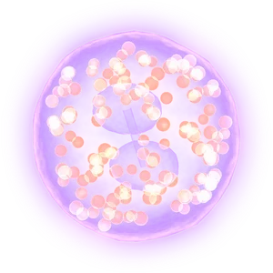
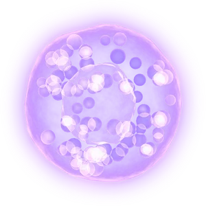
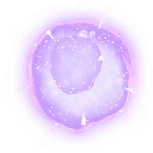
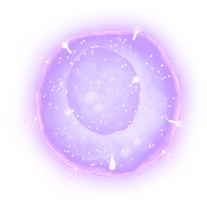
Every immune cell type carries a distinct DNA methylation signature. By quantifying immune cells by these epigenetic markers, which remain stable in dried, fixed, and frozen samples, Epiontis ID® removes the dependency on intact cells that make traditional immunophenotyping methods complex to scale as studies progress.

A novel method forged at Precision for Medicine, proven in 235+ clinical studies.


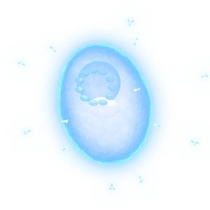
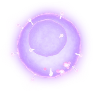
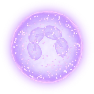
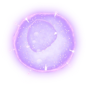
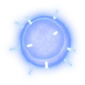
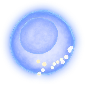
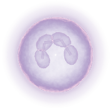
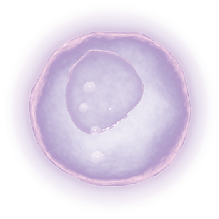
  • See the cells measured
    fig_image_111_2008981549
    sample-collection

    For diverse sample matrices​

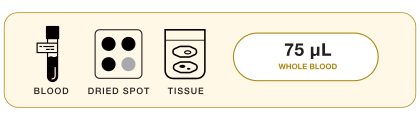
    As long as cells are present at collection, the signal is preserved in these samples to be analyzed without loss:​

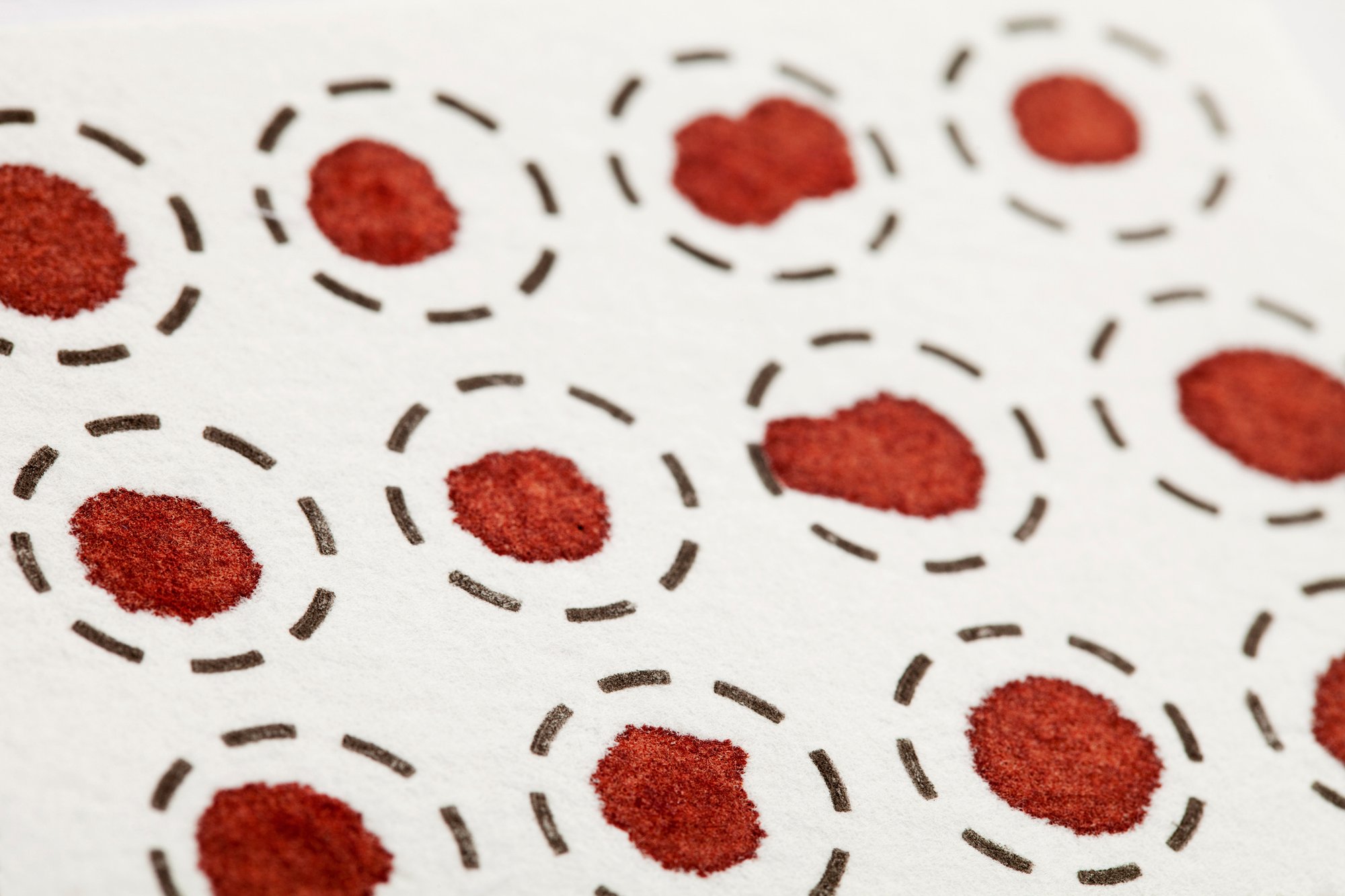
    • Dried blood spots (DBS)​

    • Biofluids​

    • FFPE tissue​

    • Fresh-frozen tissue​

    • Whole blood​

    • PBMCs​ 

    • In vitro expanded cells

    See the cells measured
    sample-collection

    For diverse sample matrices​

    As long as cells are present at collection, the signal is preserved in these samples to be analyzed without loss:​

    • Dried blood spots (DBS)​

    • Biofluids​

    • FFPE tissue​

    • Fresh-frozen tissue​

    • Whole blood​

    • PBMCs​ 

    • In vitro expanded cells

    See the cells measured
  • Pick your panel
    fig_image_145_154466925
    biomarker_labs

    For preserving timepoints without a clock​

    With no viable cell processing windows to miss, the risk of sample loss is mitigated and your immune monitoring data remains complete.​

    • DNA-based biomarkers remain stable
    • Stored samples stay useful​
    Pick your panel
    biomarker_labs

    For preserving timepoints without a clock​

    With no viable cell processing windows to miss, the risk of sample loss is mitigated and your immune monitoring data remains complete.​

    • DNA-based biomarkers remain stable
    • Stored samples stay useful​
    Pick your panel
  • fig_image_39_1836186172
    clinical_trial_support

    For cost-effective scaling to global trials​

    qPCR keeps cost per sample low as trials grow to many sites, patients, and timepoints.​

    • Lower logistics costs​

    • Ideal for sites where shipping viable cells is difficult

    • Supports decentralized sampling and patient-friendly protocols​

    Request a study consult
    clinical_trial_support

    For cost-effective scaling to global trials​

    qPCR keeps cost per sample low as trials grow to many sites, patients, and timepoints.​

    • Lower logistics costs​

    • Ideal for sites where shipping viable cells is difficult

    • Supports decentralized sampling and patient-friendly protocols​

    Request a study consult


VALIDATED BIOMARKERS

One method to measure diverse immune cell populations.​

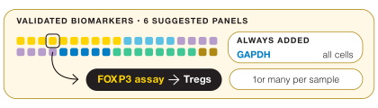
Explore the range of fully validated assays available for Epiontis ID®. Each assay targets its own methylation marker, so multiple biomarkers can be measured side by side from the same DNA, without extra blood draws or separate sample handling for each one.​

Choose a single marker, cell population, or build your own panel that covers your program’s specific immune cells of interest.​

EpiontisID®

Use arrow keys to move around a ring, Enter to select, Tab to move to the next ring.

Hover any part of the wheel. Click a cell to spin it into focus.

This explorer draws the flywheel with JavaScript.

Panel
Therapeutic area

Showing 38 of 38 validated biomarkers

Core immune population ★ Core Population

CD3+ T cells

CD3 OncAutoInfect
Learn more
CD3+ T cells
T cells & subsets · Core immune population

CD3+ T cells

Biomarker · CD3 Core assay ★ Core Population
What it is
T lymphocytes: all cells expressing the T-cell receptor–CD3 complex, the adaptive immune cells that mature in the thymus.
What it tells you
The universal T-cell denominator: nearly every other T-cell count is expressed as a share of it.
Defined by
TCR–CD3 complex on the surface
Panel · subset
T cells & subsets · Core immune population
Therapeutic areas
OncologyAutoimmune & inflammationHematologyInfectious disease
Core immune population ★ Core Population

CD4+ T cells

CD4 OncAutoInfect
Learn more
CD4+ T cells
T cells & subsets · Core immune population

CD4+ T cells

Biomarker · CD4 Core assay ★ Core Population
What it is
T cells expressing the CD4 co-receptor, which recognize antigen presented on MHC class II molecules.
What it tells you
Helper T cells that coordinate the immune response, and a population many autoimmune therapies target.
Defined by
CD4 co-receptor
Panel · subset
T cells & subsets · Core immune population
Therapeutic areas
OncologyAutoimmune & inflammationHematologyInfectious disease
Core immune population ★ Core Population

CD8+ T cells

CD8B OncAutoInfect
Learn more
CD8+ T cells
T cells & subsets · Core immune population

CD8+ T cells

Biomarker · CD8B Core assay ★ Core Population
What it is
T cells expressing the CD8 co-receptor, which recognize antigen presented on MHC class I molecules and can kill target cells.
What it tells you
Killer T cells that destroy tumor and virus-infected cells: the main effector population in immuno-oncology.
Defined by
CD8αβ co-receptor
Panel · subset
T cells & subsets · Core immune population
Therapeutic areas
OncologyAutoimmune & inflammationHematologyInfectious disease
Core immune population

Regulatory T cells (Treg)

FOXP3 OncAutoInfect
Learn more
Regulatory T cells (Treg)
T cells & subsets · Core immune population

Regulatory T cells (Treg)

Biomarker · FOXP3 Core assay
What it is
CD4+ T cells expressing FOXP3 that suppress immune responses and maintain tolerance to self.
What it tells you
The immune system’s brakes. They suppress other immune cells to prevent autoimmunity, and tumors exploit them to evade attack.
Defined by
FOXP3 transcription factor (nucleus); CD25 high, CD127 low
Panel · subset
T cells & subsets · Core immune population
Therapeutic areas
OncologyAutoimmune & inflammationHematologyInfectious disease
T-helper subsets

Th17 cells

IL17A Auto
Learn more
Th17 cells
T cells & subsets · T-helper subsets

Th17 cells

Biomarker · IL17A Core assay
What it is
CD4+ helper T cells that produce IL-17 and defend against extracellular bacteria and fungi at barrier tissues.
What it tells you
Helper T cells that make IL-17: key drivers of psoriasis, also implicated in psoriatic arthritis, axial spondyloarthritis and other autoimmune diseases.
Defined by
RORγt transcription factor; secretes IL-17
Panel · subset
T cells & subsets · T-helper subsets
Therapeutic areas
OncologyAutoimmune & inflammationHematologyInfectious disease
T-helper subsets

Tfh cells

LIF Auto
Learn more
Tfh cells
T cells & subsets · T-helper subsets

Tfh cells

Biomarker · LIF Core assay
What it is
CD4+ helper T cells that home to B-cell follicles and support germinal-center antibody responses.
What it tells you
Follicular helper T cells that help B cells form germinal centers and make high-affinity antibodies. Often expanded in lupus and Sjögren’s.
Defined by
CXCR5, PD-1, BCL6
Panel · subset
T cells & subsets · T-helper subsets
Therapeutic areas
OncologyAutoimmune & inflammationHematologyInfectious disease
T-helper subsets

Th2 cells

GATA3 AutoInfect
Learn more
Th2 cells
T cells & subsets · T-helper subsets

Th2 cells

Biomarker · GATA3 Extended assay
What it is
CD4+ helper T cells that produce IL-4, IL-5 and IL-13 and coordinate responses to parasites and allergens.
What it tells you
Helper T cells programmed by GATA3 that drive type 2 inflammation in asthma, atopic dermatitis and eosinophilic esophagitis.
Defined by
GATA3 transcription factor (nucleus); IL-4, IL-5, IL-13
Panel · subset
T cells & subsets · T-helper subsets
Therapeutic areas
OncologyAutoimmune & inflammationHematologyInfectious disease
T-helper subsets

Memory CD4+ T cells

MAF OncAutoInfect
Learn more
Memory CD4+ T cells
T cells & subsets · T-helper subsets

Memory CD4+ T cells

Biomarker · MAF Extended assay
What it is
CD4+ T cells that have previously encountered their antigen and persist long-term to respond rapidly on re-exposure.
What it tells you
Helper T cells that have met their antigen before and respond faster: a read on durability of response.
Defined by
CD45RO; antigen-experienced
Panel · subset
T cells & subsets · T-helper subsets
Therapeutic areas
OncologyAutoimmune & inflammationHematologyInfectious disease
Specialized T-cell subsets

γδ T cells

γδ T-cell assay Onc
Learn more
γδ T cells
T cells & subsets · Specialized T-cell subsets

γδ T cells

Biomarker · γδ T-cell assay Extended assay
What it is
T cells whose T-cell receptor is made of γ and δ chains instead of α and β; enriched in epithelial and mucosal tissues.
What it tells you
An unconventional T-cell subset that recognizes targets without classical MHC antigen presentation, of interest for allogeneic cell therapy.
Defined by
γδ T-cell receptor
Panel · subset
T cells & subsets · Specialized T-cell subsets
Therapeutic areas
OncologyAutoimmune & inflammationHematologyInfectious disease
Specialized T-cell subsets

Naive CD8+ T cells

CD248 Onc
Learn more
Naive CD8+ T cells
T cells & subsets · Specialized T-cell subsets

Naive CD8+ T cells

Biomarker · CD248 Extended assay
What it is
CD8+ T cells that have left the thymus but not yet encountered their specific antigen.
What it tells you
Killer T cells not yet exposed to antigen: a read on baseline T-cell fitness and apheresis starting material.
Defined by
CD45RA, CCR7, CD62L
Panel · subset
T cells & subsets · Specialized T-cell subsets
Therapeutic areas
OncologyAutoimmune & inflammationHematologyInfectious disease
Core immune population ★ Core Population

B cells

LRP5 AutoHeme
Learn more
B cells
B-cell lineage · Core immune population

B cells

Biomarker · LRP5 Core assay ★ Core Population
What it is
Lymphocytes that develop in the bone marrow, carry a B-cell receptor and can differentiate into antibody-secreting cells.
What it tells you
All B cells: the antibody-producing lineage, and the main population depleted by anti-CD20 therapies.
Defined by
CD19, CD20, B-cell receptor
Panel · subset
B-cell lineage · Core immune population
Therapeutic areas
OncologyAutoimmune & inflammationHematologyInfectious disease
B-cell subsets

Naive B cells

C7ORF50 AutoHeme
Learn more
Naive B cells
B-cell lineage · B-cell subsets

Naive B cells

Biomarker · C7ORF50 Core assay
What it is
Mature B cells that have not yet encountered their specific antigen.
What it tells you
B cells not yet exposed to antigen. Their return after depletion shows the compartment is reconstituting.
Defined by
IgD+, CD27−
Panel · subset
B-cell lineage · B-cell subsets
Therapeutic areas
OncologyAutoimmune & inflammationHematologyInfectious disease
B-cell subsets

Memory B cells

CBX6 AutoHeme
Learn more
Memory B cells
B-cell lineage · B-cell subsets

Memory B cells

Biomarker · CBX6 Core assay
What it is
B cells generated after antigen exposure that persist long-term and respond rapidly on re-exposure.
What it tells you
B cells primed by prior antigen exposure. Their return after anti-CD20 therapy can be an early marker of relapse risk.
Defined by
CD27+; mostly class-switched (IgG, IgA)
Panel · subset
B-cell lineage · B-cell subsets
Therapeutic areas
OncologyAutoimmune & inflammationHematologyInfectious disease
B-cell subsets

IgM+ B cells

IgMH AutoHeme
Learn more
IgM+ B cells
B-cell lineage · B-cell subsets

IgM+ B cells

Biomarker · IgMH Extended assay
What it is
B cells expressing IgM on their surface, including naive B cells and IgM memory B cells.
What it tells you
B cells carrying surface IgM, the first antibody class made before class switching. Relevant in CLL, Waldenström’s macroglobulinemia and class-switch defects.
Defined by
Surface IgM
Panel · subset
B-cell lineage · B-cell subsets
Therapeutic areas
OncologyAutoimmune & inflammationHematologyInfectious disease
B-cell subsets

Plasma cells

TRIO AutoHeme
Learn more
Plasma cells
B-cell lineage · B-cell subsets

Plasma cells

Biomarker · TRIO Extended assay
What it is
Terminally differentiated B cells that secrete large amounts of antibody.
What it tells you
Antibody factories: the malignant cell in multiple myeloma and the source of autoantibodies.
Defined by
CD138; secretes antibody
Panel · subset
B-cell lineage · B-cell subsets
Therapeutic areas
OncologyAutoimmune & inflammationHematologyInfectious disease
Core immune population ★ Core Population

NK cells

OSBPL5 · MVD OncHeme
Learn more
NK cells
Innate immunity & myeloid · Core immune population

NK cells

Biomarker · OSBPL5 · MVD Core assay ★ Core Population
What it is
Innate lymphocytes that kill virus-infected and tumor cells without prior sensitization.
What it tells you
Innate killers that need no prior sensitization, and the effectors behind antibody-dependent killing (ADCC).
Defined by
CD56, CD16; no CD3
Panel · subset
Innate immunity & myeloid · Core immune population
Therapeutic areas
OncologyAutoimmune & inflammationHematologyInfectious disease
Core immune population ★ Core Population

Neutrophils

LCN2 OncAuto
Learn more
Neutrophils
Innate immunity & myeloid · Core immune population

Neutrophils

Biomarker · LCN2 Core assay ★ Core Population
What it is
The most abundant circulating white blood cells: phagocytic granulocytes that are first to arrive at sites of infection.
What it tells you
The most abundant white blood cell and first responder to infection; high counts signal systemic inflammation.
Defined by
CD15, CD16
Panel · subset
Innate immunity & myeloid · Core immune population
Therapeutic areas
OncologyAutoimmune & inflammationHematologyInfectious disease
Core immune population ★ Core Population

Monocytes

PARK2 OncAuto
Learn more
Monocytes
Innate immunity & myeloid · Core immune population

Monocytes

Biomarker · PARK2 Core assay ★ Core Population
What it is
Circulating myeloid cells that can migrate into tissues and differentiate into macrophages or dendritic cells.
What it tells you
Circulating precursors of tissue macrophages and some dendritic cells: a read on myeloid burden and inflammation.
Defined by
CD14
Panel · subset
Innate immunity & myeloid · Core immune population
Therapeutic areas
OncologyAutoimmune & inflammationHematologyInfectious disease
Core immune population

Eosinophils

PRG2 Auto
Learn more
Eosinophils
Innate immunity & myeloid · Core immune population

Eosinophils

Biomarker · PRG2 Extended assay
What it is
Granulocytes containing cytotoxic granule proteins, involved in defense against parasites and in allergic inflammation.
What it tells you
Granulocytes that drive allergic, type 2 inflammation, and the cell that anti-IL-5 and anti-IL-5R therapies aim to reduce.
Defined by
Siglec-8, CCR3
Panel · subset
Innate immunity & myeloid · Core immune population
Therapeutic areas
OncologyAutoimmune & inflammationHematologyInfectious disease
Core immune population

Basophils

MCC Auto
Learn more
Basophils
Innate immunity & myeloid · Core immune population

Basophils

Biomarker · MCC Extended assay
What it is
The rarest circulating granulocytes, which release histamine and other mediators in allergic reactions.
What it tells you
The rarest granulocyte in blood, releasing histamine in allergic responses; implicated in allergy, asthma and chronic urticaria.
Defined by
FcεRI, CD123, CD203c
Panel · subset
Innate immunity & myeloid · Core immune population
Therapeutic areas
OncologyAutoimmune & inflammationHematologyInfectious disease
Monocyte & macrophage

Non-classical monocytes

C10A2213 Auto
Learn more
Non-classical monocytes
Innate immunity & myeloid · Monocyte & macrophage

Non-classical monocytes

Biomarker · C10A2213 Extended assay
What it is
A monocyte subset (CD14 low, CD16 high) that patrols the walls of blood vessels.
What it tells you
“Patrolling” monocytes that crawl along blood-vessel walls, linked to vascular and autoimmune inflammation.
Defined by
CD14 low, CD16 high
Panel · subset
Innate immunity & myeloid · Monocyte & macrophage
Therapeutic areas
OncologyAutoimmune & inflammationHematologyInfectious disease
Monocyte & macrophage

Monocytic MDSCs

ERGIC1 Onc
Learn more
Monocytic MDSCs
Innate immunity & myeloid · Monocyte & macrophage

Monocytic MDSCs

Biomarker · ERGIC1 Extended assay
What it is
Immature monocyte-like myeloid cells that suppress T-cell responses.
What it tells you
Suppressive myeloid cells tumors recruit to shut down T-cell attack, linked to checkpoint resistance.
Defined by
CD14+, HLA-DR low; suppressive function
Panel · subset
Innate immunity & myeloid · Monocyte & macrophage
Therapeutic areas
OncologyAutoimmune & inflammationHematologyInfectious disease
Dendritic cells

Plasmacytoid DCs

B9D2 OncAuto
Learn more
Plasmacytoid DCs
Innate immunity & myeloid · Dendritic cells

Plasmacytoid DCs

Biomarker · B9D2 Extended assay
What it is
Dendritic cells specialized in producing large amounts of type I interferon in response to viruses.
What it tells you
Specialized dendritic cells that produce large amounts of type I interferon: a major source of the interferon signature in lupus.
Defined by
CD123, CD303; type I interferon
Panel · subset
Innate immunity & myeloid · Dendritic cells
Therapeutic areas
OncologyAutoimmune & inflammationHematologyInfectious disease
Immune checkpoint

PD-1+ cells

PD1 OncAuto
Learn more
PD-1+ cells
Checkpoints & cytotoxic · Immune checkpoint

PD-1+ cells

Biomarker · PD1 Core assay
What it is
Immune cells, mainly activated and exhausted T cells, that express the inhibitory receptor PD-1.
What it tells you
Cells carrying PD-1, an inhibitory receptor tumors engage through PD-L1 to switch off T cells, and a major checkpoint drug target.
Defined by
PD-1 checkpoint receptor
Panel · subset
Checkpoints & cytotoxic · Immune checkpoint
Therapeutic areas
OncologyAutoimmune & inflammationHematologyInfectious disease
Immune checkpoint

TIGIT+ cells

TIGIT Onc
Learn more
TIGIT+ cells
Checkpoints & cytotoxic · Immune checkpoint

TIGIT+ cells

Biomarker · TIGIT Core assay
What it is
T cells and NK cells that express the inhibitory receptor TIGIT.
What it tells you
Cells carrying TIGIT, an inhibitory checkpoint being studied mainly in combination with anti-PD-1 therapy.
Defined by
TIGIT checkpoint receptor
Panel · subset
Checkpoints & cytotoxic · Immune checkpoint
Therapeutic areas
OncologyAutoimmune & inflammationHematologyInfectious disease
Immune checkpoint

CTLA-4+ cells

CTLA4 OncAuto
Learn more
CTLA-4+ cells
Checkpoints & cytotoxic · Immune checkpoint

CTLA-4+ cells

Biomarker · CTLA4 Core assay
What it is
T cells, especially regulatory and recently activated T cells, that express the inhibitory receptor CTLA-4.
What it tells you
Cells carrying CTLA-4, the first checkpoint targeted by an approved cancer drug. A CTLA-4 fusion protein is used to dampen T-cell activation in rheumatoid arthritis.
Defined by
CTLA-4 checkpoint receptor
Panel · subset
Checkpoints & cytotoxic · Immune checkpoint
Therapeutic areas
OncologyAutoimmune & inflammationHematologyInfectious disease
Immune checkpoint

LAG-3+ cells

LAG3 Onc
Learn more
LAG-3+ cells
Checkpoints & cytotoxic · Immune checkpoint

LAG-3+ cells

Biomarker · LAG3 Core assay
What it is
Activated T cells and some NK cells that express the inhibitory receptor LAG-3.
What it tells you
Cells carrying LAG-3, the third checkpoint with an approved therapy, used in melanoma together with anti-PD-1.
Defined by
LAG-3 checkpoint receptor
Panel · subset
Checkpoints & cytotoxic · Immune checkpoint
Therapeutic areas
OncologyAutoimmune & inflammationHematologyInfectious disease
Cytotoxic effector

Granulysin+ cells

GNLY OncAuto
Learn more
Granulysin+ cells
Checkpoints & cytotoxic · Cytotoxic effector

Granulysin+ cells

Biomarker · GNLY Extended assay
What it is
Cytotoxic T cells and NK cells that carry granulysin, an antimicrobial and cytotoxic protein.
What it tells you
Cells armed with granulysin, a cytotoxic protein: a direct read on killing activity in tumors or graft rejection.
Defined by
Granulysin in cytotoxic granules
Panel · subset
Checkpoints & cytotoxic · Cytotoxic effector
Therapeutic areas
OncologyAutoimmune & inflammationHematologyInfectious disease
Chemokine receptors

CXCR3+ cells

CXCR3 OncAuto
Learn more
CXCR3+ cells
Migration & inflammation · Chemokine receptors

CXCR3+ cells

Biomarker · CXCR3 Core assay
What it is
Immune cells, mainly Th1 cells, cytotoxic T cells and NK cells, that express the chemokine receptor CXCR3.
What it tells you
Cells carrying CXCR3, the receptor that guides Th1 and cytotoxic T cells into inflamed tissue and tumors.
Defined by
CXCR3 chemokine receptor
Panel · subset
Migration & inflammation · Chemokine receptors
Therapeutic areas
OncologyAutoimmune & inflammationHematologyInfectious disease
Chemokine receptors

CCR7+ cells

CCR7 OncHeme
Learn more
CCR7+ cells
Migration & inflammation · Chemokine receptors

CCR7+ cells

Biomarker · CCR7 Core assay
What it is
Immune cells, mainly naive and central memory T cells, that express the lymph-node homing receptor CCR7.
What it tells you
Cells carrying CCR7, which directs them into lymph nodes. Marks naive and central memory T cells.
Defined by
CCR7 lymph-node homing receptor
Panel · subset
Migration & inflammation · Chemokine receptors
Therapeutic areas
OncologyAutoimmune & inflammationHematologyInfectious disease
Chemokine receptors

CCR6+ cells

CCR6 Auto
Learn more
CCR6+ cells
Migration & inflammation · Chemokine receptors

CCR6+ cells

Biomarker · CCR6 Extended assay
What it is
Immune cells, including Th17 cells and B cells, that express the chemokine receptor CCR6.
What it tells you
Cells carrying CCR6, which guides Th17 and other cells toward inflamed skin and gut; implicated in psoriasis and IBD.
Defined by
CCR6 chemokine receptor
Panel · subset
Migration & inflammation · Chemokine receptors
Therapeutic areas
OncologyAutoimmune & inflammationHematologyInfectious disease
Chemokine receptors

CCR9+ cells

CCR9 Auto
Learn more
CCR9+ cells
Migration & inflammation · Chemokine receptors

CCR9+ cells

Biomarker · CCR9 Extended assay
What it is
Lymphocytes that express the chemokine receptor CCR9, which directs them to the small intestine.
What it tells you
Cells carrying CCR9, the gut-homing receptor that directs lymphocytes to the small intestine.
Defined by
CCR9 gut-homing receptor
Panel · subset
Migration & inflammation · Chemokine receptors
Therapeutic areas
OncologyAutoimmune & inflammationHematologyInfectious disease
Activation & migration

S1PR1+ cells

S1PR1 Auto
Learn more
S1PR1+ cells
Migration & inflammation · Activation & migration

S1PR1+ cells

Biomarker · S1PR1 Core assay
What it is
Lymphocytes that express sphingosine-1-phosphate receptor 1, which they need to exit lymph nodes.
What it tells you
Lymphocytes need S1P receptor 1 to leave lymph nodes: the target of S1P-receptor modulators in multiple sclerosis and ulcerative colitis.
Defined by
S1P receptor 1 (lymph-node exit)
Panel · subset
Migration & inflammation · Activation & migration
Therapeutic areas
OncologyAutoimmune & inflammationHematologyInfectious disease
Activation & migration

S1PR5+ cells

S1PR5 Auto
Learn more
S1PR5+ cells
Migration & inflammation · Activation & migration

S1PR5+ cells

Biomarker · S1PR5 Core assay
What it is
NK cells and some effector T cells that express sphingosine-1-phosphate receptor 5.
What it tells you
S1P receptor 5 helps NK cells leave the bone marrow and lymph nodes, and is also targeted by some S1P-receptor modulators.
Defined by
S1P receptor 5
Panel · subset
Migration & inflammation · Activation & migration
Therapeutic areas
OncologyAutoimmune & inflammationHematologyInfectious disease
Receptor & inflammation

Integrin α4+ cells

ITGA4 Auto
Learn more
Integrin α4+ cells
Migration & inflammation · Receptor & inflammation

Integrin α4+ cells

Biomarker · ITGA4 Extended assay
What it is
Leukocytes that express integrin α4, an adhesion molecule used to enter tissues such as the brain and gut.
What it tells you
The adhesion molecule that lets lymphocytes cross into the brain and gut, blocked by anti-integrin therapies in MS and IBD.
Defined by
Integrin α4 (α4β1 / α4β7)
Panel · subset
Migration & inflammation · Receptor & inflammation
Therapeutic areas
OncologyAutoimmune & inflammationHematologyInfectious disease
Receptor & inflammation

IL-6R+ cells

IL6R AutoHeme
Learn more
IL-6R+ cells
Migration & inflammation · Receptor & inflammation

IL-6R+ cells

Biomarker · IL6R Extended assay
What it is
Cells that express the IL-6 receptor, including monocytes, neutrophils and some T cells.
What it tells you
Cells carrying the IL-6 receptor, a key inflammatory signal targeted in rheumatoid arthritis and in CAR-T cytokine release syndrome.
Defined by
IL-6 receptor
Panel · subset
Migration & inflammation · Receptor & inflammation
Therapeutic areas
OncologyAutoimmune & inflammationHematologyInfectious disease
Fibrosis & structural

COL1A1+ cells

COL1A1 OncAuto
Learn more
COL1A1+ cells
Non-leukocyte populations · Fibrosis & structural

COL1A1+ cells

Biomarker · COL1A1 Core assay
What it is
Fibroblasts that produce type I collagen, the main structural protein of connective tissue and scar.
What it tells you
Fibroblasts actively producing type I collagen, laying down scar tissue in lung, liver and skin fibrosis.
Defined by
Producing type I collagen
Panel · subset
Non-leukocyte populations · Fibrosis & structural
Therapeutic areas
OncologyAutoimmune & inflammationHematologyInfectious disease
Fibrosis & structural

PDGFRB+ cells

PDGFRB OncAuto
Learn more
PDGFRB+ cells
Non-leukocyte populations · Fibrosis & structural

PDGFRB+ cells

Biomarker · PDGFRB Core assay
What it is
Pericytes and fibroblasts that express PDGF receptor β, a growth-factor receptor that drives their proliferation and migration.
What it tells you
Pericytes and activated fibroblasts that contribute to scar formation in fibrosis and to dense tumor stroma.
Defined by
PDGFRβ receptor
Panel · subset
Non-leukocyte populations · Fibrosis & structural
Therapeutic areas
OncologyAutoimmune & inflammationHematologyInfectious disease

Therapeutic areas: Onc = Oncology · Auto = Autoimmune & inflammation · Heme = Hematology · Infect = Infectious disease.
★ Core Population: the seven main populations of the Immune Cell Census, to start every program. Each card is one validated assay, named for the gene at its marker region.

NOT SURE WHERE TO START?

Begin with the Immune Cell Census: Seven counts for a validated foundation​

Before adding specific subsets, every study can benefit from a baseline view of the immune system and how it shifts with disease and treatment. The Immune Cell Census provides that foundation, measuring the seven major immune populations tracked in nearly every study.​

fig_image_111_2008981549

CD3+ T cells
CD3

fig_image_111_2008981549

CD4+ T cells
CD4

fig_image_111_2008981549

CD8+ T cells
CD8B

fig_form_raster_131_197042805

B cells
LRP5

fig_image_143_499213572

NK cells
0SBPL5 - MVD

fig_image_144_63895775

Neutrophils
LCN2

fig_image_145_154466925
Monocytes
PARK2

POPULATION COUNTED
ASSAY - MARKER REGION
WHAT IT TELLS YOU
CD3+ T cells CD3 Total T cells: the denominator for every T-cell subset
CD4+ T cells CD4 Helper T cell
CD8+ T cells CD8B Cytotoxic T cells
B cells LRP5 B-cell depletion and recovery on B-cell-directed therapies
NK cells 0SBPL5 blood - MVD tissue Innate cytotoxic cells
Neutrophils LCN2 The most abundant circulating leukocytes; inflammation
Monocytes PARK2 Circulating myeloid cells, precursors of macrophages

HOW IT WORKS​

Counted by DNA, not by living cells.​

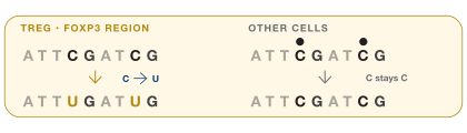
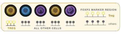
Epiontis ID® identifies immune cell populations through lineage-specific methylation markers written in DNA. This enables precise immune cell quantification – both relative and absolute counts – that correlates closely with flow, while minimal processing and standardized analysis support comparable results across sites and over time.​

Example cell populations and associated gene markers methylated CpG demethylated CpG = Epiontis ID
CHROMOSOME X156 Mb · drawn to scale p q Xp11.23 FOXP3· 14.3 kb chrX:49,250,438 49,264,710 marker region

Gene

FOXP3forkhead box P3
XChromosome
Xp11.23Cytogenetic band
14.3 kbGene size

chrX:49,250,438–49,264,710 · GRCh38

Encodes a forkhead/winged-helix transcription factor that programs regulatory T-cell development and suppressive function. Mutations cause IPEX syndrome.

Associated Cell Measurement

Regulatory T cells

CD4+ T cells expressing FOXP3 that suppress immune responses and maintain tolerance to self.

1 ID per cell type. A marker region demethylated only in that cell type: its epigenetic ID.

Gene locations: NCBI Gene / GRCh38. The marker region’s position inside the gene is illustrative; chromosome banding is schematic.

One ID per cell type​

Reads the marker region demethylated only in that cell type: its unique epigenetic ID.​

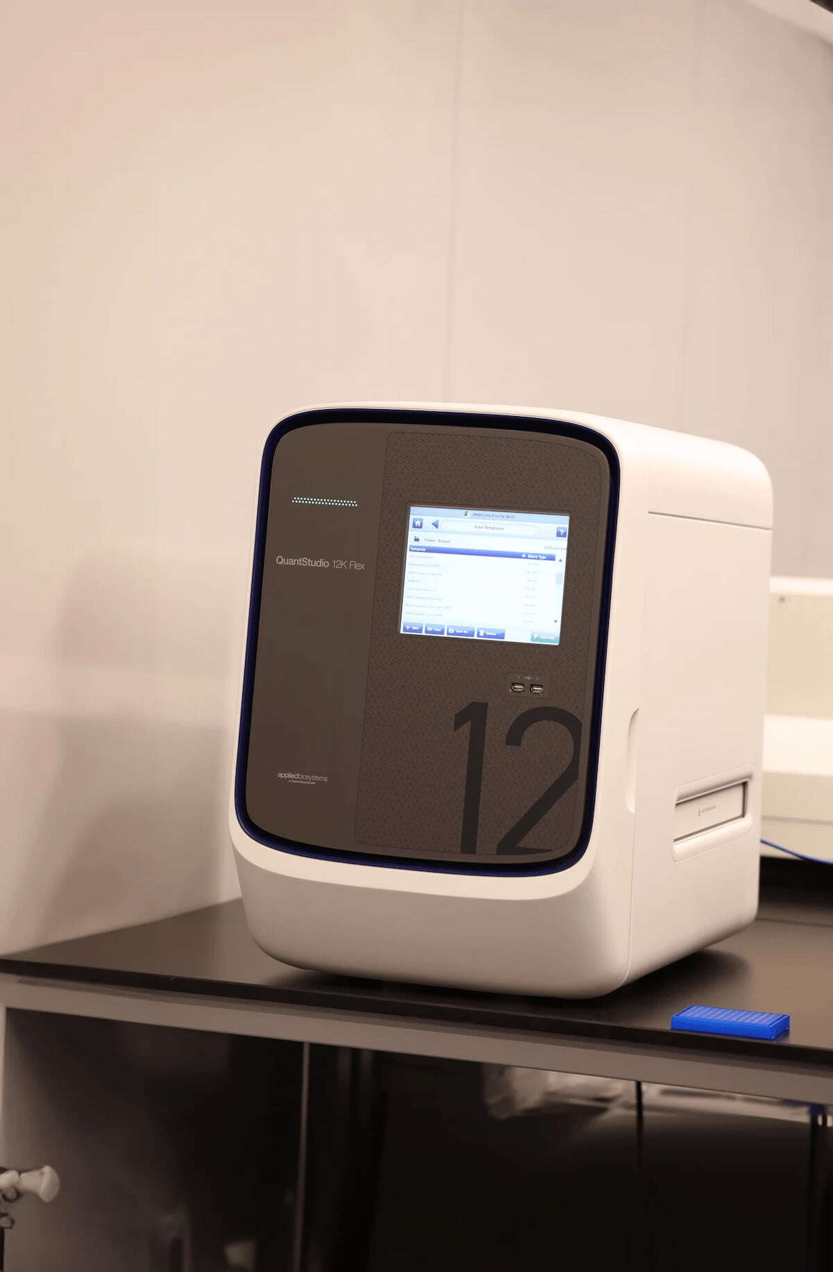
qPCR readout​

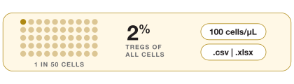
Digital, normalized, and automated for relative (% of all cells) and absolute (cells/uL) counts.​


Epiontis ID®, Flow Cytometry, or Both?​

Ensure the right readout for your sample, phase, and scientific question.

Some studies need the depth of flow cytometry, others need the reach of Epiontis ID®, and many need both.​

Brochure

Epiontis ID: A Different Approach to Immune Monitoring

Download Brochure
Epiontis ID: A Different Approach to Immune Monitoring

From sample to cell count in seven steps.​

  • 1.

    Collect a sample

    Whole blood, a dried blood spot or tissue: frozen, ambient or fixed. As little as 75 µL of blood.

    PfM_EpiontisID_Immune Identity_P8_Step 1


  • 2.

    Choose the biomarkers

    Each biomarker is one assay, with primers for one cell type’s marker region. Choosing the assay defines the question.

    PfM_EpiontisID_Immune Identity_P8_Step 2


  • 3.

    Bisulfite conversion

    Unmethylated C becomes U (read as T). Methylated C is protected. An invisible tag becomes a sequence difference.

    PfM_EpiontisID_Immune Identity_P8_Step 3


  • 4.

    Extract DNA from all cells

    Cells don’t need to be alive. The methylation pattern was set during differentiation; the assay only reads it.

    PfM_EpiontisID_Immune Identity_P8_Step 4


  • 5.

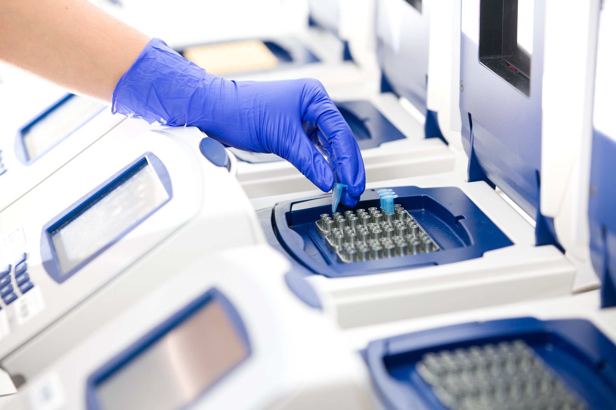
    Methylation-specific qPCR

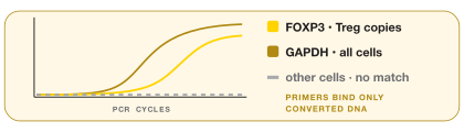
    Primers match only the converted, demethylated marker region, so only the target cell type is amplified.

    PfM_EpiontisID_Immune Identity_P8_Step 5


  • 6.

    Normalize

    GAPDH, demethylated in every cell, counts all cells. A plasmid µL standard, added at the start, corrects PCR efficiency.

    PfM_EpiontisID_Immune Identity_P8_Step 5


  • 7.

    Report

    Each population as % of total cells and,  for blood, cells/µL, in your desired report format, with an optional bioanalytical report.

    PfM_EpiontisID_Immune Identity_P8_Step 7


  • Dried Blood Sample
  • fig_image_2_55
  • 22.-Gene-mutation,-damage-to-a-part-of-a-cells-DNA
  • DNA-1
  • iStock-177381768
  • EOS52022-1
  • PQI Internal KOM- Report banners 2025 GTR_Header
A PRECISION FOR MEDICINE INNOVATION

Expanding the possibilities for immune monitoring across therapeutic areas, with trusted clinical results.

Epionits ID® was developed by Precision for Medicine scientists to measure what traditional markers could not always distinguish: a cell’s true lineage identity. This DNA methylation-based approach has evolved into a scalable platform for targeted immune monitoring, trusted by 17 of the top 20 pharma companies and used in 235+ clinical studies and counting.​

  • Brochure

    Epiontis ID: Use Cases Across Diverse Therapeutic Areas

    Download Brochure
    Epiontis ID: Use Cases Across Diverse Therapeutic Areas
  • Case Study

    Use of Epiontis ID® to support understanding therapeutic efficacy in a phase 2 psoriasis trial

    Download Case Study
    Use of Epiontis ID® to support understanding therapeutic efficacy in a phase 2 psoriasis trial

WHY EPIONTIS ID®?

Epigenetic immune monitoring that is affordable to start and built to scale.​

Epiontis ID® expands what is possible for immunophenotyping beyond viable cell workflows – offering  an alternative or orthogonal approach for:​

  • Large, global trials and remote sites with complex logistics​

  • Studies where intact cells are not available or hard to obtain​

  • Obtaining immune data on known target cells, cost-effectively

fig_container_raster_72_789517194

 
Delivered by Precision for Medicine’s expert team.​

Our global specialty labs, sample processing facilities and lab partners bring one standardized Epiontis ID® method to sites across North America, Europe and Asia Pacific, with kitting, logistics and sample management built in.​

Explore our integrated approach >>


Frequently Asked Questions

Does Epiontis ID® replace flow cytometry?

In some cases, yes. Epiontis ID complements flow cytometry, is an alternative in certain circumstances, and can be used where flow isn’t feasible, such as with frozen, dried or fixed samples. In whole blood, results have been shown to correlate well.

How much sample is needed?

As little as 75 µL of whole blood covers a panel of four cell types. Dried blood spots and tissue also work.

Can you measure in tissue?

Yes. Many assays are validated in FFPE and  fresh-frozen tissue; coverage varies by assay.

What if the population we need isn’t on the list?

New marker regions can be co-developed, typically in 12–18 months. CAR T-cell programs are one example.

How do Epiontis ID® costs compare to flow cytometry?

Epiontis ID measurements are economically priced, with additional budget savings: overnight shipping and site-level preparation of individual samples are not needed.
In fact, average savings on international shipping costs alone can be reduced by as much as 92%.*

*Internally verified calculations for Epiontis ID using dried blood spot vs. ambient blood on a global scale

How many assays per sample?

Each biomarker is its own assay. Cost effective panel designs include 4, 9, or 14 assays per sample. Any number of assays can be included in your panel, in any combination.

Can samples collected in China be analyzed?

Yes, in-country through a partner third-party laboratory, with data returned for one consistent report.

Is this approach scientifically validated?

Yes. The approach is supported by hundreds of peer-reviewed publications. For regulatory T cells, Nobel laureate Shimon Sakaguchi has referenced Precision for Medicine’s work on the epigenetic regulation of Tregs.