Quantitative Multiplex Immunofluorescence
Multiplex immunofluorescence (IF) image analysis can be an invaluable tool in immuno-oncology and autoimmune indications or where an understanding of the status of the immune system is important.

Single-cell and spatial biology insights with multiplex immunofluorescence
For multiplex IF assays, Precision uses instruments including the PhenoImager HT (previously known as the Vectra® Polaris™) Automated Quantitative Pathology Imaging System by Akoya, which is a robust and flexible platform that can be used in a CLIA setting and is compatible with both tissues and cells.
We offer assay development and validation of multiplex IF staining using multiple types of multispectral image detection technologies. For studies requiring higher plex, we also support Akoya PhenoCycler-Fusion workflows to analyze dozens of proteins per slide using iterative cycling.
Precision for Medicine is also recognized as a Certified Partner of Akoya Biosciences for multiplex immunofluorescence tissue profiling, demonstrating our dedication to using advanced technologies for precise and efficient tissue profiling.
A selection of capabilities Precision provides using mIF
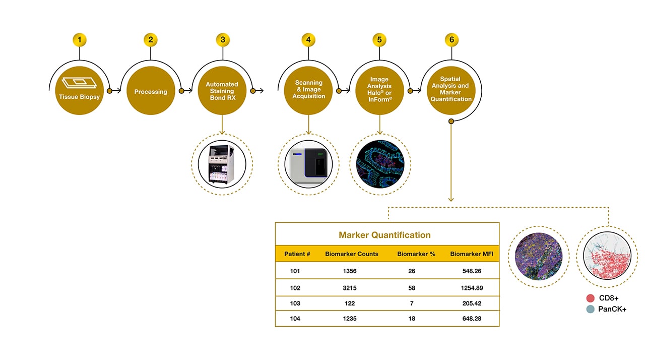
Precision’s mIF workflow can detect and visualize up to 8 markers of interest simultaneously from a sample, and enable panels of 60+ markers for whole-slide phenotyping. This generates complex spatial information of a tumor or tissue sample, and can be used to visualize the spatial distribution of proteins within a single cell—all of which can provide information on pharmacodynamics (PD), mechanism of action, and progression or state of disease. Precision offers quantitative and high-throughput multiplex IF on fresh and archival tissues as well as cells isolated from liquid biopsies using ApoStream®, such as circulating tumor cells (CTCs).
AI-powered image analysis can be performed using Inform or Halo patented software to visualize, analyze, quantify, and phenotype abundant and rare cells, including subcellular localization (nuclear vs cytoplasmic) of proteins at the single cell level. We also offer tiered assay qualification and validation up to CLIA-regulated use.
- Phenotype cells within a sample
- Nearest neighbor analysis
- Infiltration analysis
- Analyze individual cells
-
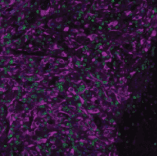
Using 9-color imaging, phenotype all cells from a sample into predefined subpopulations.
Shown is an example of an ovarian cancer tumor biopsy, with single-cell intensity and spatial distribution. Each individual cell from the whole slide scan has been classified into a predefined phenotype.

-
Nearest neighbor analysis quantifies cell-cell interactions by measuring the distance between each cell of one phenotype (phenotype A) and its closest neighbor of another phenotype (phenotype B). This analysis helps quantify spatial relationships critical for understanding immune-tumor dynamics.
The distance between phenotype A to the closest neighbor of phenotype B can be used to generate plots and average differences in mean distance when comparing pretreatment and posttreatment samples.

-
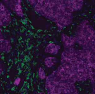
Infiltration analysis quantifies the number, penetration, and migration of immune cell infiltration and can be used to quantify any number of predefined phenotypes located within a specified distance from a predefined annotation.
In this analysis, the red line corresponds to the tumor margin, drawn by AI-assisted manual annotation. The dotted red, pink, and green lines are software-generated concentric bins each representing approximately 50 μm in thickness.

-
Biomarkers in individual cells can be measured — for example in circulating tumor cells (CTCs). Precision's CTC capture technology, ApoStream®, can be utilized to collect and concentrate CTCs, and pre-qualified mIF panels targeted to a selection of different cancer types can then deliver cytology information. This approach supports exploratory and translational oncology studies.

Target the clinical biomarkers important in your trials
See which biomarkers we've worked with and some we've for immunochemistry and multiple immunofluorescence.

Whitepaper: The role of the tumor microenvironment, and how quantitative mIF can be leveraged for tumor profiling
Quantitative Multiplex Immunofluorescence: A Powerful Tool for Oncology Therapeutic Development
Download Whitepaper
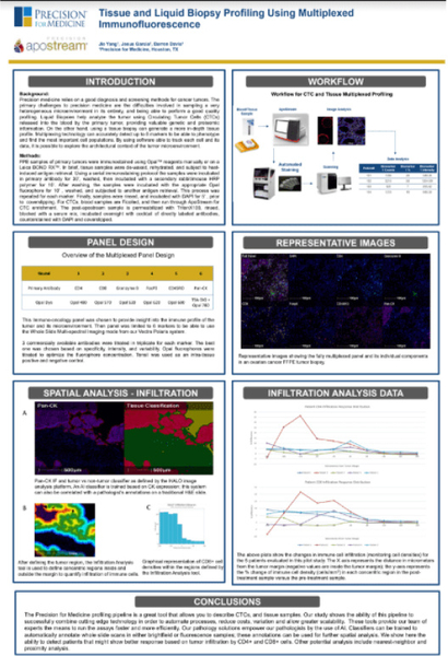
Poster: Tissue and Liquid Biopsy Profiling Using Multiplexed Immunofluorescence
In this poster, we demonstrate how multiplex IF can be used to effectively explore the architectural context of the tumor microenvironment and classify tumors as hot, cold, or immune-ignored.
Related specialty lab services
-
Explore


IHC
End-to-end IHC solutions including kitting, sample logistics, staining via all major platforms, and in-house pathology services
IHC
End-to-end IHC solutions including kitting, sample logistics, staining via all major platforms, and in-house pathology services -
Explore


ApoStream®: CTC Liquid Biopsy
Proprietary Precision technology for antibody-free isolation and concentration of target cells in liquid biopsy samples; enables analysis via methods including mIF, FISH, and NGS
ApoStream®: CTC Liquid Biopsy
Proprietary Precision technology for antibody-free isolation and concentration of target cells in liquid biopsy samples; enables analysis via methods including mIF, FISH, and NGS -
Explore


Genomics
With NGS, NanoString, qPCR and ddPCR capabilities, we can add insights from genomic and transcriptomic technologies to your understanding of patient biology.
Genomics
With NGS, NanoString, qPCR and ddPCR capabilities, we can add insights from genomic and transcriptomic technologies to your understanding of patient biology.