Polyamines (PAs) are involved in cell growth, proliferation, migration and gene regulation, and the maintenance of cellular oxidative homeostasis.1 Deregulation of PA metabolism has been associated with various epithelial cancers and neurodegenerative diseases. Alterations in PA metabolism may cause cellular damage and apoptosis through the generation of oxidative byproducts of PA catabolism, which occurs via two pathways. One of these pathways involves the enzyme spermine oxidase (SMOX), which converts spermine to spermidine and generates reactive hydrogen peroxide and aldehydes as byproducts.
There is a growing body of evidence linking SMOX activity with DNA damage, inflammation, and carcinogenesis, garnering interest in this enzyme as a potential drug target. Increased expression of this enzyme is frequently found in epithelial cancer models and studies in in vivo models have shown that treatment with a polyamine oxidase inhibitor lowers spermidine levels, decreases DNA damage and inflammation, and reduces tumor numbers. However, research has been hampered by the lack of well-characterized, validated antibodies for identifying target human SMOX (hSMOX) proteins.
An evaluation of three commercially available affinity-purified polyclonal antibody preparations found that these preparations lacked the sensitivity and specificity required for supporting downstream applications.2 To address this challenge, an industry sponsor sought to develop a panel of specific and selective rabbit monoclonal antibodies against various hSMOX epitopes for different applications, including immunohistochemistry (IHC) based analyses of human cancer tissues.
Development and Characterization of Rabbit Monoclonal Antibodies
The industry sponsor selected five different immunization peptides, guided by a publicly available hSMOX homology model. Rabbits were immunized with each of the five peptides and full-length recombinant hSMOX (rhSMOX). After ELISA screening, 24 clones, referred to as SMABs, were selected for larger scale mAb expression and purification.
Epitope binning experiments were performed with 16 purified SMABs to establish which of them bind to epitopes in close proximity. In addition, activity assays measuring the production of hydrogen peroxide were performed to identify which SMABs inhibit the enzymatic activity of rhSMOX. To select SMABs for in-depth performance testing using human tissues, western blot and immunofluorescence staining were performed.
IHC to evaluate Human Cancer Tissues
Note: Precision for Medicine developed and optimized the IHC assays and performed all IHC experiments and tissue work in the evaluation performed.
Initial assay optimization
For optimization of IHC conditions to detect hSMOX using SMAB antibodies, negative (Raji cells), low- (A549 cells) and high-expressing (stimulated A549 cells) cell line controls were chosen based on existing literature and western blot and immunofluorescence results.
To serve as a model for tissue staining, the cell lines were processed to formalin-fixed paraffin-embedded (FFPE) blocks. From these, a cell tissue microarray (SMOX-TMA) was constructed, and sections were used for the selection of SMAB antibodies based on their performance in IHC (see Figure 1).

From the SMABs evaluated, SMAB10 and SMAB17 demonstrated a desirable profile with no staining in the negative cell line control and a clear contrast in staining between low- and high-expressing A549 cell controls.
Further assay optimization
Given that SMAB10 showed slightly better specificity, an IHC assay using SMAB10 was further optimized using the SMOX-TMA. This SMAB10 IHC assay was also used to assess immunoreactivity in tumor xenografts comprising the

Assay qualification
Qualification of the SMAB10 IHC assay protocol for human tissues staining was performed on nine full-face tumor sections, including colon, stomach, lung, liver, pancreas and prostate carcinomas.
For each IHC assay, human normal colon FFPE sections were prepared and stained using anti-von Willebrand Factor (anti-vWF) antibody as an assay control sample to validate the detection reagents. IHC was performed using a Dako PLUS autostainer and EnVision FLEX Kit reagents. Sections were incubated with peroxidase blocker and then serum-free protein block prior to incubation with anti-SMOX primary antibodies applied at a range of concentrations. Following application of EnVision FLEX HRP-Polymer, sections were incubated with DAB HRP-substrate prior to counterstaining with hematoxylin, dehydration and coverslipping. Finally, the slides were scanned using Leica Biosystems’ Aperio AT turbo scanner.
Antibody qualification
To validate the use of SMAB10 antibody to detect SMOX in normal and cancer human samples, the qualified IHC conditions for SMAB10 were applied to multi-tumor TMAs surveying samples of four common human malignancies and reference normal tissues.
Pathologist review
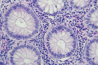
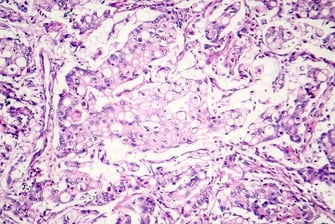
SMAB10 staining in human tumor TMAs was binned into four classes based on semi-quantitative immunohistochemical intensity scoring (negative or grade 1-3). This scoring assessment was performed by visual inspection by one of Precision for Medicine’s board-certified pathologists. This analysis confirmed that SMOX is expressed more frequently in multiple cancers compared to non-diseased tissues, with the greatest intensity and highest frequency of positive hSMOX staining in colon cancer tissue. Positive tumor staining was also visible in breast and prostate cancer tissue and was least frequent and intense in lung cancer tissues.

(A) hSMOX staining on human tumor TMAs using a SMAB10 IHC assay. (B). Grading of hSMOX staining frequency in human tumor TMAs.
Conclusion
The availability of specific, selective and high-affinity monoclonal antibodies is essential for visualizing, detecting, and quantifying hSMOX in different experimental settings and applications. In this study, researchers generated and characterized a series of new monoclonal antibodies that recognize hSMOX as a tool for studying SMOX biology and facilitating SMOX drug development. To visualize hSMOX in different cell lines or human tissues, Precision for Medicine delivered the development, optimization, and validation of an IHC assay utilizing SMAB10 and used that assay to demonstrate that SMAB10 staining appears more frequently in cancer tissue compared to normal tissue, supporting a potential role for SMOX in cancer development.
Explore our extensive IHC capabilities and expert pathology services >
References
1. Damiani E, Wallace HM. Polyamines and cancer. Methods Mol Biol. 2018;1694:469–488.
2. Tepper AWJW, et al. Development and characterization of rabbit monoclonal antibodies that recognize human spermine oxidase and application to immunohistochemistry of human cancer tissues. PLoS One. 2022;17(4):e0267046.